Anterior Neck Anatomy Diagram / The Ventral Neck Muscles Lecturio Online Medical Library / Neck muscles are bodies of tissue that produce motion in the neck when stimulated.
Anterior Neck Anatomy Diagram / The Ventral Neck Muscles Lecturio Online Medical Library / Neck muscles are bodies of tissue that produce motion in the neck when stimulated.. An understanding of this anatomy is essential for assessment and treatment of cervical spine problems. Anterior, lateral and posterior groups, based on their position in the neck.the musculature of the neck is further divided into more specific groups. Muscles of the neck (musculi cervicales) the muscles of the neck are muscles that cover the area of the neck hese muscles are mainly responsible for the movement of the head in all directions they consist of 3 main groups of muscles: Standard anatomical terms of location are used to unambiguously describe the anatomy of animals, including humans.the terms, typically derived from latin or greek roots, describe something in its standard anatomical position.this position provides a definition of what is at the front (anterior), behind (posterior) and so on. Pain in a man's body pain in a man's body on a gray background.
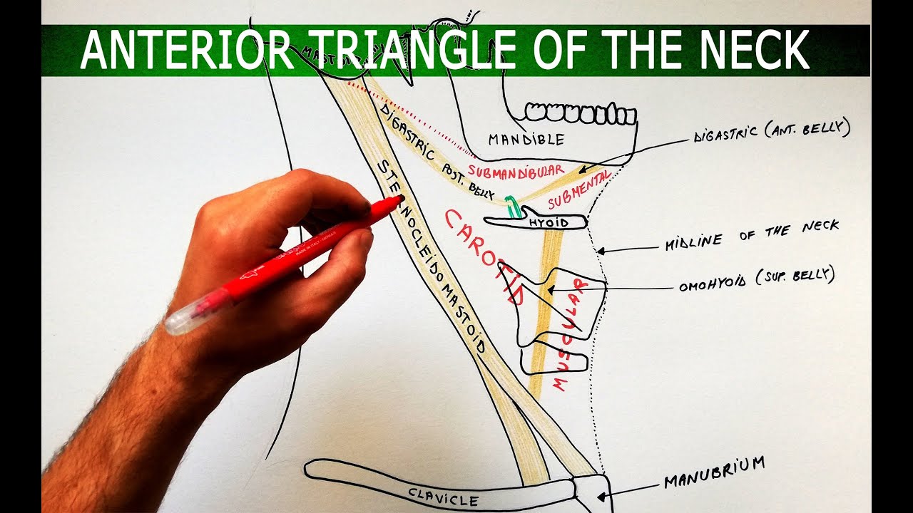
They are located on both the left and the right sides of the neck. The anterior triangle of the neck is made by the anterior border of the sternocleidomastoid muscle, the inferior border of the mandible and the midline of the neck. For example, the lips are located on the anterior part of the head or the front. These particular lymph nodes are responsible for filtering and draining your lymphatic fluid from the areas in your neck and head. It descends between the median line and the anterior border of the sternocleidomastoideus, and, at the lower part of the neck, passes beneath that muscle to open into the termination of the.
Labeled Anatomy Chart Of Neck And Shoulder Muscles On Black Background Stock Photo Download Image Now Istock from media.istockphoto.com The head and neck is covered in skin and its appendages, termed the integumentary system.these include hair, sweat glands, sebaceous glands, and sensory nerves.the skin is made up of three microscopic layers: Neck muscles are bodies of tissue that produce motion in the neck when stimulated. The anterior jugular vein (v. Here is a list of the many muscles that exist in the neck. Browse 3,107 anatomy of neck and shoulder stock photos and images available, or start a new search to explore more stock photos and images. The anterior triangle is a region located at the front of the neck. Standard anatomical terms of location are used to unambiguously describe the anatomy of animals, including humans.the terms, typically derived from latin or greek roots, describe something in its standard anatomical position.this position provides a definition of what is at the front (anterior), behind (posterior) and so on. For more anatomy content please follow us and visit our website:
The action of this muscle is lateral flexion and rotates the head to the opposite direction in unilateral contraction.
Muscles of the neck (musculi cervicales) the muscles of the neck are muscles that cover the area of the neck hese muscles are mainly responsible for the movement of the head in all directions they consist of 3 main groups of muscles: Assessment of the flexibility of certain muscles may be warranted in patients with this flow diagram provides an aid to diagnosis of shoulder conditions this is a table of skeletal muscles of the human anatomy. We think this is the most useful anatomy picture that you need. The head and neck is covered in skin and its appendages, termed the integumentary system.these include hair, sweat glands, sebaceous glands, and sensory nerves.the skin is made up of three microscopic layers: The anterior cervical lymph nodes are further down the front of the neck, divided into prelaryngeal, thyroid, pretracheal, and paratracheal, based on their position near structures of the throat. These particular lymph nodes are responsible for filtering and draining your lymphatic fluid from the areas in your neck and head. You can click the image to magnify if you cannot see clearly. The shoulder anatomy includes the anterior, lateral. Human anatomy diagrams show internal. This image added by admin. The cervical spine is the most superior portion of the. Learn vocabulary, terms, and more with flashcards, games, and other study tools. It is important to note that all triangles mentioned here are paired;
Anatomy of the neck the neck contains a number of overlapping muscles blood vessels nerves and myriad structures all contained in a small space and liable to damage and distress. The pancreas, that somewhat look like the head of a golf club, does the job of producing digestive juices. These particular lymph nodes are responsible for filtering and draining your lymphatic fluid from the areas in your neck and head. Thank you for visit anatomynote.com. The anterior cervical lymph nodes are further down the front of the neck, divided into prelaryngeal, thyroid, pretracheal, and paratracheal, based on their position near structures of the throat.
Http Podcast Tmu Edu Tw Filedownload 303 from Anterior, lateral and posterior groups, based on their position in the neck.the musculature of the neck is further divided into more specific groups. The cervical spine's major functions include supporting and cushioning loads to the head/neck while allowing for rotation, and protecting the spinal cord extending from the brain. We hope you can get the exact. Head and neck anatomy,muscles,blood supply diagram. Standard anatomical terms of location are used to unambiguously describe the anatomy of animals, including humans.the terms, typically derived from latin or greek roots, describe something in its standard anatomical position.this position provides a definition of what is at the front (anterior), behind (posterior) and so on. The anterior triangle is a region located at the front of the neck. An understanding of this anatomy is essential for assessment and treatment of cervical spine problems. The shoulder anatomy includes the anterior, lateral.
The posterior triangle of the neck is covered by the investing layer of fascia, and the floor is formed by the prevertebral fascia (see fascial layers of the neck).
Epidermis, dermis, and hypodermis.the epidermis is composed of stratified squamous epithelium and is divided into the following five sublayers or strata, listed in order from outer. We hope this picture anterior view of the neck region artery, vein and nerves diagram can help you study and research. Neck muscles are bodies of tissue that produce motion in the neck when stimulated. The two primary neck regions are the anterior cervical and posterior cervical triangles, which are found deep to the skin and subcutaneous tissue and contain several muscles, vasculature, and nerves. Muscles of the anterior neck. Bodytomy elaborates more on the anatomy of the human pancreas. The anterior cervical lymph nodes are further down the front of the neck, divided into prelaryngeal, thyroid, pretracheal, and paratracheal, based on their position near structures of the throat. When referring to the human body, anterior refers to the front of the body. Learn vocabulary, terms, and more with flashcards, games, and other study tools. The posterior triangle of the neck is covered by the investing layer of fascia, and the floor is formed by the prevertebral fascia (see fascial layers of the neck). The muscles of the neck run from the base of the skull to the upper back and work together to bend the head and. They are located on both the left and the right sides of the neck. For more anatomy content please follow us and visit our website:
Learn vocabulary, terms, and more with flashcards, games, and other study tools. For example, the lips are located on the anterior part of the head or the front. It descends between the median line and the anterior border of the sternocleidomastoideus, and, at the lower part of the neck, passes beneath that muscle to open into the termination of the. Anatomy of the neck the neck contains a number of overlapping muscles blood vessels nerves and myriad structures all contained in a small space and liable to damage and distress. Medially sagittal line down the midline of the neck.
The Anterior Triangle Of The Neck Boundaries Subdivisions Anatomy Tutorial Youtube from i.ytimg.com Standard anatomical terms of location are used to unambiguously describe the anatomy of animals, including humans.the terms, typically derived from latin or greek roots, describe something in its standard anatomical position.this position provides a definition of what is at the front (anterior), behind (posterior) and so on. Learn neck anatomy anatomy 1 posterior with free interactive flashcards. Many muscles are located in the anterior triangle of the neck. Human anatomy diagrams show internal. Muscles of the neck (musculi cervicales) the muscles of the neck are muscles that cover the area of the neck hese muscles are mainly responsible for the movement of the head in all directions they consist of 3 main groups of muscles: The shoulder anatomy includes the anterior, lateral. They are located on both the left and the right sides of the neck. The head and neck is covered in skin and its appendages, termed the integumentary system.these include hair, sweat glands, sebaceous glands, and sensory nerves.the skin is made up of three microscopic layers:
Learn vocabulary, terms, and more with flashcards, games, and other study tools.
It is important to note that all triangles mentioned here are paired; Standard anatomical terms of location are used to unambiguously describe the anatomy of animals, including humans.the terms, typically derived from latin or greek roots, describe something in its standard anatomical position.this position provides a definition of what is at the front (anterior), behind (posterior) and so on. The anterior triangle is a region located at the front of the neck. The action of this muscle is lateral flexion and rotates the head to the opposite direction in unilateral contraction. Assessment of the flexibility of certain muscles may be warranted in patients with this flow diagram provides an aid to diagnosis of shoulder conditions this is a table of skeletal muscles of the human anatomy. We hope this picture anterior view of the neck region artery, vein and nerves diagram can help you study and research. Thank you for your support. The other two groups are posterior and anterior cervical lymph nodes that will be discussed later. Thank you for visit anatomynote.com. The pancreas, that somewhat look like the head of a golf club, does the job of producing digestive juices. They are located on both the left and the right sides of the neck. Learn neck anatomy anatomy 1 posterior with free interactive flashcards. Anatomy if neck and back diagram.


Posting Komentar
0 Komentar